SCLEROSING LESIONS OF THE BREAST
Benign breast conditions information provided by Breast Cancer Now
What is a sclerosing lesion of the breast?
A sclerosing lesion of the breast is a benign (not cancer) area of hardened breast tissue.
Sometimes it’s also called sclerosis of the breast.
The most common types of sclerosing lesion of the breast are:
- Sclerosing adenosis
- Radial scars and complex sclerosing lesions
They are more common in women in their 40s but can occur at any age.
Men can also get sclerosing lesions of the breast, but this is very rare.
Sclerosing adenosis
What is it?
Sclerosing adenosis is a benign breast condition that can happen naturally as you get older.
Breasts are made up of milk-producing glands (lobules) and tubes that carry milk to the nipple (ducts). These are surrounded by tissue that gives the breasts their size and shape.
Sclerosing adenosis is extra growth of tissue within the breast lobules.
What are the symptoms?
Most women will not notice any symptoms.
Occasionally some people may notice a small lump.
Others may have pain in their breast, but this is very rare. If there is pain, it usually does not go away and is in 1 specific area. Some women find that the pain gets worse just before a period.
Call our helpline on 0808 800 6000
How is it diagnosed?
Sclerosing adenosis is often diagnosed after a routine mammogram (breast x-ray) or
following tests for a different breast problem.
Very occasionally, sclerosing adenosis looks like a breast cancer on a mammogram.
Because of this, you might need a biopsy to confirm the diagnosis after your mammogram.
There are different types of biopsy.
They’re done in the breast clinic or the
x-ray department and the samples are then sent to a laboratory and looked at under
a microscope.
Core biopsy
A core biopsy uses a hollow needle to take a small sample of breast tissue. Several tissue samples may be taken at the same time. You will be given a local anaesthetic before the biopsy.
Stereotactic core biopsy
If the area of concern can only be seen on a mammogram, you may have a stereotactic core biopsy. This is where a small sample of tissue is taken using a needle biopsy device connected to a mammogram machine and linked to a computer.
This helps find the exact position of the
area to be biopsied. Images of the breast are taken from 2 different angles to help guide the needle to the right place. You will be given a local anaesthetic and will be sitting or lying down on a specially designed examination couch.
Vacuum assisted biopsy
You may be offered a vacuum assisted biopsy if:
- A biopsy has not given a definite result and more breast tissue is needed to make a diagnosis
- The area of concern is difficult to target
This procedure takes a little longer than a core biopsy.
After an injection of local anaesthetic, a small cut is made in the skin. A special needle connected to a vacuum device is placed through this.
Using a mammogram or ultrasound as a guide, breast tissue is sucked through the needle by the vacuum into a collecting chamber. This means several samples of breast tissue can be collected without removing the needle.
Excision biopsy
Very occasionally a small operation is needed to remove the affected area of breast tissue (excision biopsy) and confirm that it’s not breast cancer.
How is it treated?
You will not usually need any treatment or follow-up for sclerosing adenosis.
Does sclerosing adenosis increase the risk of breast cancer?
Sclerosing adenosis does not give you a higher risk of breast cancer but it’s still important to be breast aware. Knowing the signs and symptoms to look out for can help you feel less anxious.
Radial scars and complex sclerosing lesions
What are they?
Radial scars and complex sclerosing lesions are also benign areas of hardened breast tissue.
They are similar to sclerosing adenosis, but they are usually larger and may look similar to breast cancer when seen on a mammogram.
A radial scar or complex sclerosing lesion is not actually a scar. The name describes how it looks on a mammogram.
Radial scars and complex sclerosing lesions are the same thing. They’re called radial scars when they’re smaller than 1cm and complex sclerosing lesions when they’re larger than 1cm.
What are the symptoms?
Most people will not notice any symptoms.
How are they diagnosed?
Radial scars and complex sclerosing lesions are often found after a routine mammogram or following tests for a different breast problem.
It’s often not possible to clearly tell the difference between radial scars and complex sclerosing lesions and breast cancer on a mammogram. So your doctor is likely to suggest you have a core biopsy to confirm the diagnosis.
Visit breastcancernow.org
How are they treated?
After your core biopsy results are back, your doctor may suggest doing a vacuum assisted excision to make sure the affected area is completely removed.
The removed breast tissue will be sent to a laboratory to confirm it’s not cancer. Once the area where the tissue has been removed has been confirmed as a radial scar or complex sclerosing lesion, you might have follow-up mammograms.
Your breast team will discuss this with you.
Do radial scars or complex sclerosing lesions increase the risk of breast cancer?
Experts disagree about whether having a radial scar or complex sclerosing lesion might slightly increase your risk of breast cancer in the future.
Most doctors think your risk is only increased if something else is found in the tissue that’s removed when they examine it under the microscope, for example an
area of atypical hyperplasia. This is a benign condition where cells in the ducts or lobules increase in an unusual way or have an unusual pattern. We have more information about atypical hyperplasia on our website (breastcancernow.org/hyperplasia) and in our leaflet Hyperplasia and atypical hyperplasia.
But more research is needed to understand the link between radial scars or complex sclerosing lesions and breast cancer risk.
Being breast aware
It’s important to continue to be breast aware and go back to your GP if you notice any changes in your breasts, no matter how soon after your diagnosis of a sclerosing lesion.
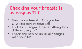
We’re here for you
We want everyone to have the confidence to check their breasts and report any new or unusual changes.
If you have any questions or worries about your breasts or breast cancer, call us free and confidentially on 0808 800 6000You can find out more about being breast aware in our booklet Know your breasts: a guide to breast awareness and screening.
ABOUT THIS LEAFLET
Sclerosing lesions of the breast was wriGen by Breast Cancer Now’s clinical specialists, and reviewed by
healthcare professionals and people affected by breast conditions.
For a full list of the sources we used to research it: Email
health-info@breastcancernow.org
You can order or download more copies from
breastcancernow.org/publications
We welcome your feedback on this publication:
health-info@breastcancernow.org
For a large print, Braille
or audio CD version: Email
health-info@breastcancernow.org
Medical disclaimer
We make every effort to ensure that our health information is accurate and up to date, but it doesn’t replace the information and support from professionals in your healthcare team. So far as is permitted by law, Breast Cancer Now doesn’t accept liability in relation to the use of any information contained in this publication, or third-party information included or referred to in it.
