BREAST CALCIFICATIONS
Benign breast conditions information provided by Breast Cancer Now
Causes of breast calcifications
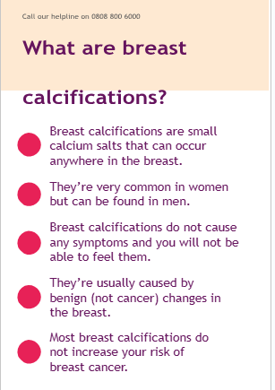
Breast calcifications are usually due to benign changes in the breast that happen as you get older.
Sometimes they form because of other benign conditions, such as a fibroadenoma or breast cyst. See our leaflets on these conditions for more information.
They can also form if you have:
- Had an infection in your breast
- Injured your breast
- Had any type of breast surgery
Breast calcifications can develop in the blood vessels and skin of the breast. These may be age- related or caused by other medical conditions.
Occasionally, breast calcifications can be an early sign of breast cancer. Because of this, you may need tests to check what sort of calcifications you have.
Diagnosing breast calcifications
Breast calcifications are usually found by chance on a routine screening mammogram or an investigation at a breast clinic for another breast problem.
Calcifications show up on a mammogram as bright white dots or specks.
Calcifications are classified as looking benign (not cancer), indeterminate (uncertain), or suspicious of being cancer based on their size, shape and pattern.
Types of breast calcifications
Microcalcifications
Microcalcifications are very small. They often occur because of benign changes, but
occasionally microcalcifications can be an early sign of cancer.
Macrocalcifications
Macrocalcifications are larger. They usually occur because of benign changes and do not need to be investigated.
Further tests
If the calcifications look benign, you will not usually need any more tests.
If the calcifications look indeterminate (uncertain) or suspicious, you’ll need more tests to help make an accurate diagnosis.
Mammogram
You may have another mammogram, called tomosynthesis, that gives a more detailed picture of the affected area.
Ultrasound scan
An ultrasound scan uses sound waves to produce an image of the breast tissue.
Core biopsy
A core biopsy uses a hollow needle to take a small sample of breast tissue. It’s done using a mammogram or ultrasound for guidance.
Several tissue samples may be taken at the same time. This procedure will be done using a local anaesthetic, where the area is numbed.
The sample will be sent to a laboratory to be looked at under a microscope.
Vacuum assisted biopsy
This procedure takes a little longer than a core biopsy.
After an injection of local anaesthetic, a small cut is made in the skin. A special needle connected to a vacuum device is placed through this.
Using a mammogram or ultrasound as a guide, breast tissue is then sucked through the needle by the vacuum into a collecting chamber. This means several samples of breast tissue can be collected without removing the needle.
Visit breastcancernow.org
The samples are sent to a laboratory to be
examined under a microscope.
Vacuum assisted excision biopsy Occasionally, you may have a vacuum assisted excision biopsy to remove an area of calcification.
This is a similar procedure to a vacuum assisted biopsy, but more tissue may be removed.
This may mean you can avoid having an operation under a general anaesthetic.
Inserting a metal marker Sometimes a small metal clip (or marker) may be placed in the breast where the
biopsy has been taken. This is so the area can easily be found again if you need another biopsy or surgery. The clip can be safely left in the breast.
The marker clip is usually made of titanium, the same metal used for joint replacement surgery. It will not set off alarms at airports.
Most clips are now suitable for having an MRI. But if the marker clip is left in and you need to have an MRI scan in the future, let your doctor or radiographer know.
Treatment
Breast calcifications do not usually
need to be treated
In most cases you will not need any treatment for breast calcifications.
If the calcifications look benign, they do not need to be removed and will not cause you any harm.
Surgery
You may need an operation to remove the area of calcification if:
- It’s not possible to get a biopsy of the area
- A biopsy does not confirm a diagnosis
- Biopsy results show an unusual change
(called atypia)
- Biopsy results show a sign of an early cancer
The operation is done under general anaesthetic, where you’re given medication to make you sleep. You can usually go home on the same day.
Identifying the area to be removed
As calcifications cannot be felt, before the operation the exact position has to be “located” so the surgeon can remove the right area.
This is called localisation. It’s done using local anaesthetic in the x-ray department, with mammogram or ultrasound as a guide.
A very small magnetic or radioactive “seed” is
inserted into the breast using a fine needle.
This can be done up to 30 days before your surgery.
During surgery, a special probe is used to locate the seed and guide the surgeon to the tissue that needs to be removed. The seed will be removed during the operation.
Sometimes a fine wire may be used instead of a seed. This is usually done on the same day as your operation and the wire removed during surgery.
AŁer surgery
After your operation, you may feel soreness and discomfort, which you can manage with pain relief.
There will be a scar, but this should fade in time.
Follow-up
If tests show the calcifications are due to benign changes, you will not need any more treatment or follow-up.
If the calcifications are part of another benign breast condition or an unusual change (atypia), you’ll be told if you need any follow-up.
If you’re told the calcifications are an early sign of breast cancer, your treatment team will discuss whether you need more tests and what the next steps are.
Do breast calcifications increase the risk of breast cancer?
Most breast calcifications do not increase your risk of breast cancer.
If the breast calcifications are due to an unusual change, this may slightly increase your risk of breast cancer. Your treatment team can discuss this further with you.
It’s important to continue to be breast aware and go back to your GP if you notice any changes in your breasts, no matter how soon after you were told you have calcifications.You can find out more about being breast aware in our booklet Know your breasts: a guide to breast awareness and screening.
ABOUT THIS LEAFLET
Breast calcifications was wriGen by Breast Cancer Now’s clinical
specialists, and reviewed by healthcare professionals and people affected
by breast conditions.
For a full list of the sources
we used to research it: Email health- info@breastcancernow.org
You can order or download more copies from
breastcancernow.org/publications
We welcome your feedback on this publication:
health-info@breastcancernow.org
For a large print, Braille
or audio CD version: Email
health-info@breastcancernow.org
Medical disclaimer
We make every effort to ensure that our health information is accurate and up to date, but it doesn’t replace the information and support from professionals in your healthcare team. So far as is permitted by law, Breast Cancer Now doesn’t accept liability in relation to the use of any information contained in this publication, or third-party information included or referred to in it
